Small Molecules: Therapeutic Application in Neuropsychiatric and Neurodegenerative Disorders

Department of Clinical and Experimental Medicine, University of Foggia, Via Napoli, 20, 71122 Foggia, Italy
*
Author to whom correspondence should be addressed.
Molecules 2018, 23(2), 411; https://doi.org/10.3390/molecules23020411
Submission received: 29 December 2017
/
Revised: 5 February 2018
/
Accepted: 7 February 2018
/
Published: 13 February 2018
(This article belongs to the Section Bioorganic Chemistry)
Abstract
:In recent years, an increasing number of studies have been published, focusing on the potential therapeutic use of small catalytic agents with strong biological properties. So far, most of these works have only regarded specific clinical fields, such as oncology, infectivology and general pathology, in particular with respect to the treatment of significant inflammatory processes. However, interesting data on possible therapeutic applications of small molecules for the treatment of neuropsychiatric and neurodegenerative illnesses are emerging, especially with respect to the possibility to modulate the cellular redox state. Indeed, a crucial role of redox dysregulation in the pathogenesis of these disorders has been widely demonstrated by both pre-clinical and clinical studies, being the reduction of the total amount of free radicals a promising novel therapeutic approach for these diseases. In this review, we focused our interest on studies published during the last ten years reporting therapeutic potential of small molecules for the treatment of neuropsychiatric and neurodegenerative disorders, also based on the biological efficiency of these compounds in detecting intracellular disturbances induced by increased production of reactive oxygen species.
1. Aims and Methodology for Literature Search
Small molecules are crucial players in several chemical reactions, able to detect the presence of a copious number of cellular metabolites [1]. Indeed, in a large variety of pathological conditions, several molecular alterations, which do not generally occur in physiological states, are detectable and mainly regard the increased production of free radicals, pH alterations and impaired generation of some specific biomolecules [2]. In recent years, an increasing number of studies focused on the potential therapeutic use of small agents with strong biological properties have been published [3], but, so far, most of the reports on this subject have mainly regarded proliferative or infectious diseases [4,5,6,7,8,9,10,11,12,13,14,15,16].
Here, we aimed to review studies published during the last ten years (from 1 November 2007 to 1 November 2017), focused on the potential therapeutic use of small agents for the treatment of neuropsychiatric and neurodegenerative disorders, in particular with respect to their bioactivity of sensoring intracellular alterations caused by enhanced free radical amount, discussing the benefits and the limits of this therapeutic approach.
The literature source for this review included open access papers found in Pubmed in the above-mentioned period using, as key words, the term small molecules in combination with the following words: neuropsychiatric disorders, neurodegenerative diseases, schizophrenia, mood disorders, anxiety, autism, Alzheimer’s disease, Parkinson’s disease, amyotrophic lateral sclerosis, stroke, spinal cord injury, traumatic brain injury. Each of these combinations has been further associated with the terms: oxidative stress, redox modulation, ROS, free radicals. By using these keyword combinations, we obtained a total number of 158 papers, with 14 duplicate records, which were removed for further screening. We then excluded 42 papers which were not written in English or were other than original research papers or reviews. A second round of exclusion concerned papers (29) which did not provide a detailed description of the chemical characteristics of the considered small molecules. Finally, 73 papers were included in the analysis. The total number of references of this paper also included the ones used for the introducing statements of the different sections of the manuscript.
2. The Use of Small Molecules in Neuropsychiatric Disorders
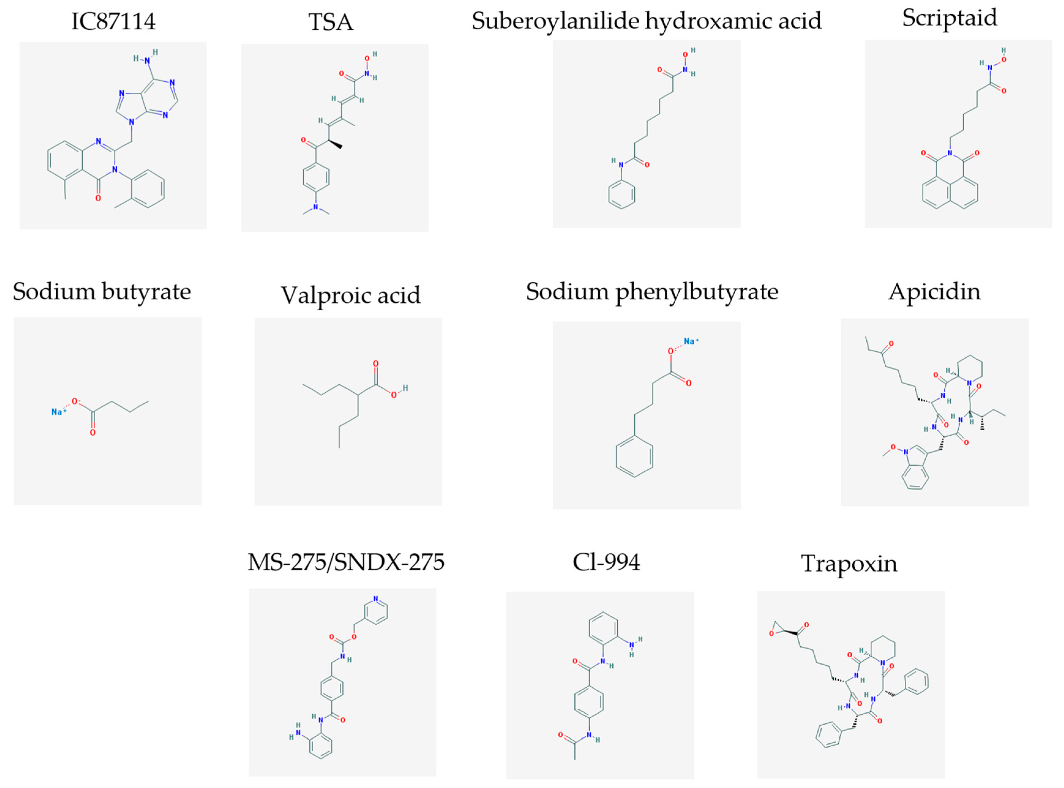
The main findings related to the possible therapeutic potential of small molecules in the treatment of schizophrenia, mood disorders, anxiety and autism are reported in Table 1. The chemical structures (https://pubchem.ncbi.nlm.nih.gov/) [17] of specific small molecules with therapeutic potential in neuropsychiatric disorders are shown in Figure 1.
3. The Use of Small Molecules in Neurodegenerative Disorders
3.1. Therapeutic Potential for Alzheimer’s Disease
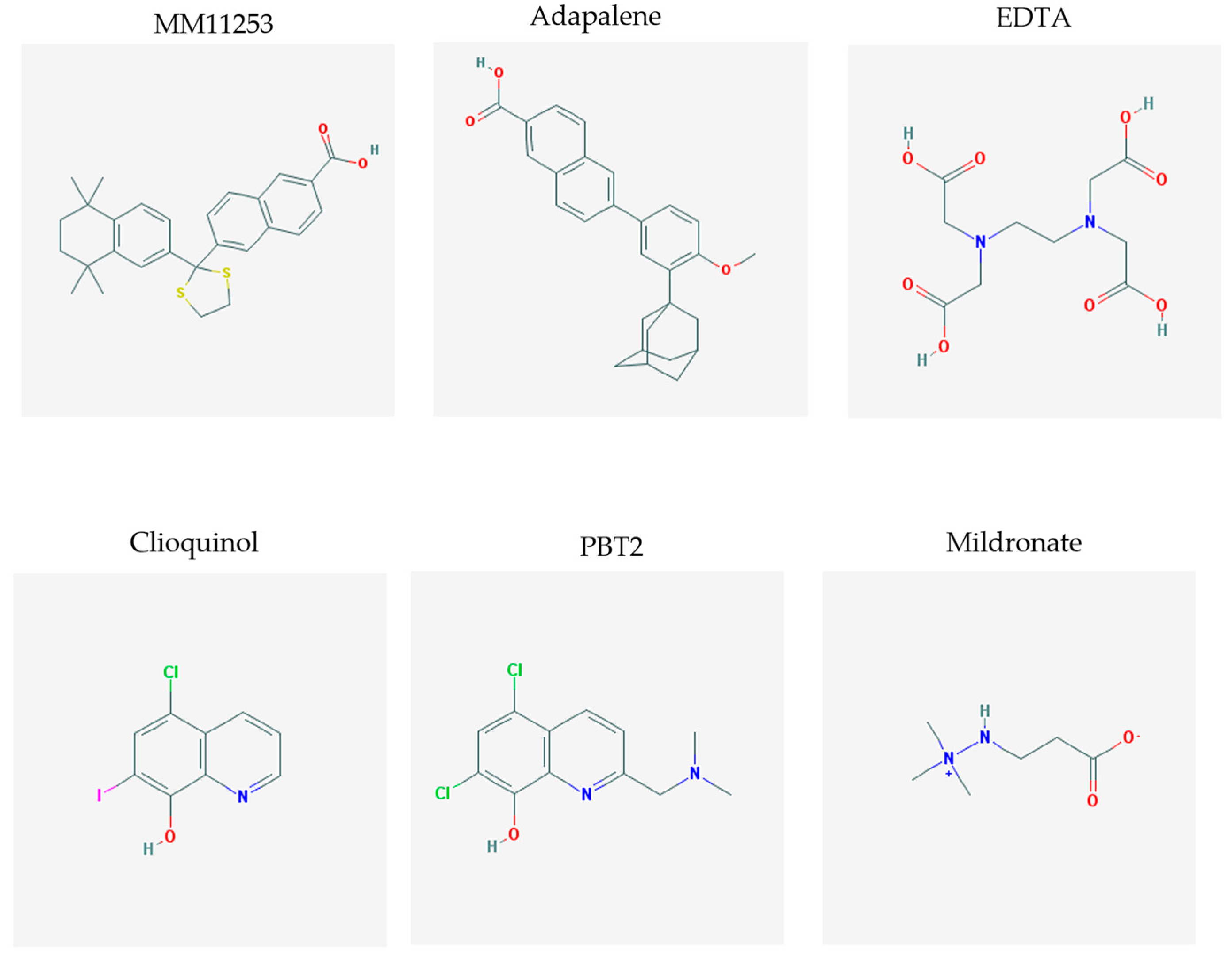
A crucial event in the pathogenesis of Alzheimer’s disease (AD) is represented by the amyloid-β peptide (Aβ) aggregation which initiates a cascade of molecular pathways, finally resulting in neuronal death and degeneration [28]. Importantly, abnormal Aβ metabolism can be detected several years before AD onset [29] and this aspect represents an important pharmacological target for early therapeutic interventions. However, so far, no compound specifically targeting the process of Aβ accumulation, and mainly developed by using animal models of the disease, has been translated into clinical practice [30]. Therefore, the identification of small molecules, acting on Aβ accumulation and aggregation, has represented the focus of an increasing number of studies in this field, in the perspective of opening a novel and promising “chapter” in the history of compounds to be used in AD. One of the most complete and straightforward works in this sense is represented by a very recent paper by Habchi and co-workers, in which the authors reported the systematic development of some small molecules, classified as “set A” (seven molecules showing a similar or greater effect than the bexarotene) and “set B” (five molecules able to totally inhibit Aβ42 aggregation for a period of at least 10 h), that inhibit specific steps of Aβ42 aggregation [31]. However, for the sake of clarity, it should be specified that some of the small molecules identified in this work, such as MM11253 and adapalene, were previously described by other authors to significantly impact not only the onset of the aggregation but also Aβ42 oligomer proliferation [32,33].
The other records found with our research strategy, highlight, rather, the development of small molecules targeting another crucial pathogenetic process in AD progression, i.e., the loss of metal ion homeostasis and their impaired compartmentalization [34,35], especially concerning the redox active Fe(II/III) and Cu(I/II), that have been found as highly concentrated in senile plaques [36,37], in the cortex and hippocampus [38,39]. Hence, these reactive metals can bind to Aβ species, then undergoing Fenton reaction, resulting in the production of specific reactive oxygen species (ROS), such as hydrogen peroxide and hydroxyl radical, which may facilitate Aβ aggregation and trigger neurodegeneration [35]. Therefore, a valuable therapeutic strategy to reduce neurotoxicity, induced by the interaction between metals and Aβ species, and to re-establish metal ion homeostasis in the brain, might be represented by the metal-Aβ association outbreak via metal chelation. So far, the most used chelators in AD therapy included ethylenediaminetetraacetic acid (EDTA), clioquinol and PBT2, an 8-hydroxyquinoline derivative. In the attempt to translate preclinical findings to possible clinical application, the last two compounds were also tested in phase II clinical trials and they have been reported to significantly improve cognitive functions [40,41], despite their serious side effects, such as the subacute myelo-optic neuropathy induced by clioquinol. Consistent efforts have been also addressed to the development of small molecules, such ascyclen, KLVFF peptide, curcumin, IMPY, and p-I-stilbene that were able to synergistically link both metal ions and Aβ [42,43,44], in order to overcome the limit of the previously developed metal chelating compounds, especially related to blood-brain barrier permeability.
3.2. Therapeutic Potential for Parkinson’s Disease
Parkinson’s disease (PD) is a neurodegenerative disorder characterized by the loss of the dopaminergic neurons in the substantia nigra. Several pathogenetic mechanisms have been proposed for this Central nervous system (CNS) disorder. One of the most investigated is related to mitochondrial and redox dysfunctions [45,46], including mutations in the mitochondria-specific kinase PTEN-induced kinase 1 (PINK1), as well as in the E3 ubiquitin ligase Parkin, a mitochondria-associated protein [47]. In line with this concept, several papers reported the therapeutic potential of PINK1/Parkin pathway activation in PD [48,49]. However, despite the widely known mechanism of small molecule-mediated activation of kinases, typically accomplished by binding their allosteric regulatory sites, PINK1 has been shown to not contain small molecule-binding sites, being, therefore, its pharmacological activation mediated by other mechanisms [49]. On the other hand, mildronate [3-(2,2,2-trimethylhydrazinium) propionate dihydrate], a small molecule with charged nitrogen and oxygen atoms that protect mitochondria, has been described to act as neuroprotective compound in a mouse model of neurotoxicity induced by azidothymidine, via suppression of brain inflammation and apoptosis, as well as decrease of cytochrome oxidase c, caspase-3, inducible Nitric Oxide Synthase (iNOS) and cellular apoptosis susceptibility-protein [50]. The mildronate-related neuroprotection was further confirmed in a study performed by using a rat model of PD that was obtained by unilateral intra-striatal injection of the neurotoxin 6-hydroxydopamine (6-OHDA). Indeed, mildronate administration to 6-OHDA-injected animals prevented the loss of specific biomarkers, such as tyrosine hydroxylase, ubiquitin and Notch-3, which are known to assure neural and glial integrity, simultaneously decreasing the expression of specific markers of inflammation, such as iNOS [51]. In line with mitochondria dysfunctions, another crucial pathogenetic mechanism associated to the development of PD is related, at molecular levels, to dysfunctions of GTPases, especially the ones regulating the dynamic processes of mitochondria fission and fusion, the organelle transport along axons, the axon maintenance, as well as the neuronal survival, and to neuroinflammation and oxidative stress enhancement [52]. With respect to redox imbalance, the small GTPase Rac1, which crucially regulates the functioning of the free radical producer NOX1, has been reported to accumulate in dopaminergic neurons of patients affected by PD [53]. Interestingly, a library containing 5 million small molecules has been probed as modulator for different GTPases, such as Rab5, Rab7, Cdc42, wild type Ras and mutant Ras, Rho, and Rac that selectively activate GTPase subfamilies [52].
Another significant acquired knowledge in the field of molecular mechanisms leading to PD, deals with the role of specific proteins, including α-synuclein. Indeed, in a very interesting paper of Misook and co-workers, authors attempted to synthesize a library of small molecules, in order to rapidly and efficiently identify potential pharmacological “chaperones”, i.e., small molecules that bind proteins and stabilize them against degradation, as well as novel chaperone inhibitors against α-synuclein [54].
The chemical structures (https://pubchem.ncbi.nlm.nih.gov/) [17] of small molecules with therapeutic potential in AD and PD are shown in Figure 2.
3.3. Therapeutic Potential for Amyotrophic Lateral Sclerosis
Amyotrophic lateral sclerosis (ALS) is a fatal neurodegenerative disease, characterized by motor neuron death and rapid progression, for which there are no effective therapeutic opportunities. From a pathogenetic point of view, 20% of the familiar forms of ALS are caused by mutations in the SOD1 gene [55] which induce a gain of toxic functions by motor neurons. The exact cause of ALS is still unknown. However, cell autonomous and non-cell autonomous mechanisms are reported to contribute to the degenerative process [56,57,58]. More recent research has identified TAR-DNA binding protein-43 (TDP-43) as a crucial player in the pathogenesis of both sporadic and non-SOD1 familial ALS [59,60]. Furthermore, several pathogenic initiators of ALS, including oxidative stress, mitochondrial dysfunctions, neuroinflammation and depletion of neurotrophins have been identified [61]. Despite the limited therapeutic efficacy of the current available treatments, in recent years a significant number of studies described the development and/or proposed the use of different small molecules for ALS (Table 2).
4. The Use of Small Molecules in Neurological Disorders Associated with Neurodegeneration
4.1. Therapeutic Potential for Stroke
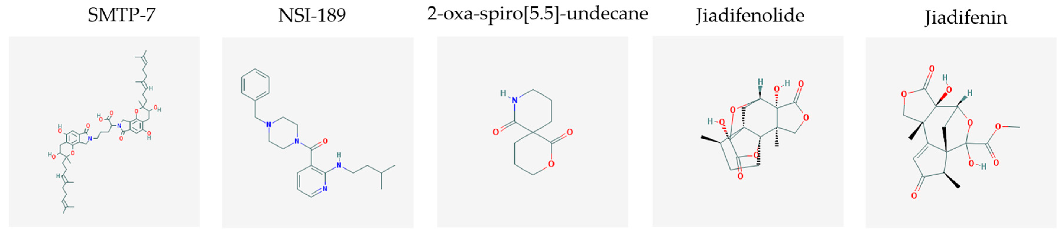
Stroke is a neurological disorder commonly associated with a very high mortality and disability rate, for which the only existing therapeutic opportunity consists in the use of a thrombolytic agent in order to restore the blood flow in the ischemic area [76]. However, only a limited number of subjects can benefit from this pharmacological intervention. Therefore, there is an urgent need to develop new therapeutic strategies. In this context, the potential use of small molecules, as possible pharmacological choice, has become a field of lively scientific research. In particular, the therapeutic effects of the small molecule Stachybotrys microspora triprenyl phenol-7 (SMTP-7), that promotes activation of plasminogen through the modulation of plasminogen conformation [77], against cerebral ischemia has been tested in several rodent models of stroke [78,79,80]. Importantly, together with its strong thrombolytic properties, SMTP-7 has been also described as a decrease of neuroinflammation related to stroke-induced neurodegeneration [78,81]. Despite the utility of data obtained on rodent models of stroke regarding this small molecule, studies in higher-order species, such as primates, whose cerebral vascularization and vessel morphology show more significant similarities with the human counterpart than rodents [82], can be considered extremely significant for a more direct translation towards the human pathology and for a potential clinical application of SMTP-7. In this perspective, the study conducted by Sawada and co-workers appears of great importance. Indeed, authors succeeded in demonstrating in primates, by using the photochemically induced thrombotic middle cerebral artery occlusion model, causing cyclic flow reductions typical of infarct progression in stroke patients, that SMTP-7 reduced cerebral infarction, neurologic deficits, and hemorrhage in the infarct area, thus also displaying a significant neuroprotection [83]. Neuroprotective effects of small molecules against stroke-associated neurodegeneration, especially in terms of neurostructural benefits and enhancement of neurogenesis, have been also clearly reported in a recent elegant work that evaluated the effects of an oral administration of the small molecule NSI-189, which is already in clinical trial for the treatment of major depression and prevention against suicide (https://clinicaltrials.gov/) [84], in the middle cerebral artery occlusion mouse model [85]. Thanks to a well-conceived experimental approach, authors were able to demonstrate that NSI-189 promoted behavioral recovery, enhanced cell proliferation and neurogenesis, and upregulated specific neurogenic factors, such as BDNF, if administered at a wider therapeutic window of 6 hours after stroke [85]. Although no data on the effects of this small molecule on primate models of stroke are currently available, results obtained on the mouse model, could be certainly considered very promising in the perspective of approving this small agent also for the treatment of stroke in humans. Another promising category of pharmacological compounds which recently raised interest in the research of novel therapeutic options for stroke and associated neurodegeneration is represented by natural product-based small molecules with neurotrophic, neurogenic and anti-neuroinflammatory actions. Recent efforts in this direction by the group of Jhelum and collaborators led to the discovery of some novel compounds based on 2-oxa-spiro[5.5]-undecane, derived from the natural product paecilomycine A, which have been described as booster of neurite growth and neuronal regeneration, and as a neuroinflammation decreaser [86,87]. Other natural small molecules, such as jiadifenolide, jiadifenin and (1R,10S)-2-oxo-3,4-dehydro-xyneomajucin (ODNM), have been also tested in vitro for their neuritogenic and neurotrophic activity, by using primary rat hippocampal neurons and ESC-derived motor neurons [88] or PC12 cells [89]. The chemical structures (https://pubchem.ncbi.nlm.nih.gov/) [17] of small molecules with therapeutic potential for stroke-related neurodegeneration are shown in Figure 3.
4.2. Therapeutic Potential for Spinal Cord Injury
4.3. Therapeutic Potential for Traumatic Brain Injury
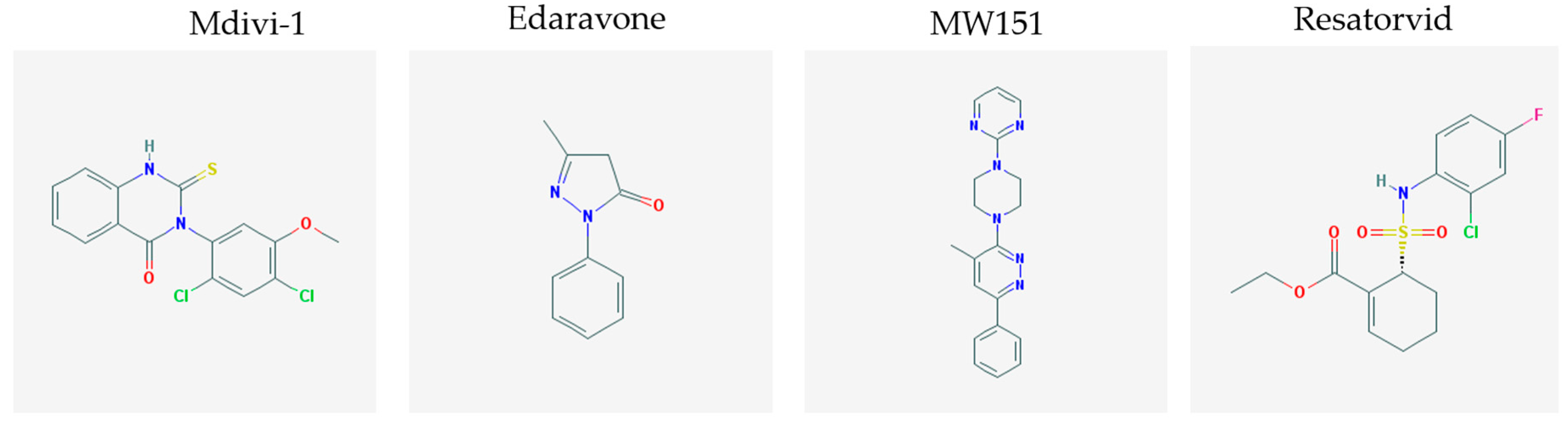
Traumatic brain injury is commonly considered an important cause of morbidity and mortality. This pathological event has been described as the initiator of a cascade of detrimental processes that can exacerbate the primary injury, worsen long-term outcome and increase the risk of neurodegenerative complications [110]. Several pathogenetic factors have been considered as crucial players in this mechanism. Among them, the most investigated one refers to mitochondria dysfunctions, with enhanced ROS generation and consequent oxidative stress and overproduction of pro-inflammatory cytokines, finally resulting in inflammation, edema, blood brain barrier loss of integrity and increased permeability, neurotoxicity, and cell death [111]. Thus, the development of small molecules that target each of these processes is the focus of an increasing interest, in order to find alternative therapeutic strategies against complications derived from neurodegeneration. With respect to mitochondria dysfunctions, a recent report of Wu and co-workers investigated the possible beneficial role of Mdivi-1, a small molecule inhibitor of the key mitochondrial fission protein dynamin-related protein 1(Drp1), against neuronal death and functional outcome deficits, in a mouse model of traumatic brain injury. By this interesting experimental approach, authors demonstrated that Drp1 inhibition, obtained by Mdivi-1 administration, significantly alleviated behavioral alterations, brain edema, impairment of mitochondrial morphology and cell death induced by traumatic brain injury [112].
Evaluation of the potential therapeutic effects of small molecules that reduce oxidative stress has been also conducted by Wang and Co-workers, who demonstrated the neuroprotective effects of edaravone, a synthetic free radical scavenger small molecule, in a rat model of traumatic brain injury. Indeed, administration of this compound 2 and 12 h after traumatic brain injury significantly decreased neuronal loss and death, and reduced oxidative stress, also acting on non-neuronal cell types, such as astrocytes and microglia. Furthermore, this compound was also shown to positively act on blood brain barrier loss of integrity and altered functioning, as well as to decrease the production of inflammatory cytokines [113].
In line with the evaluation of small molecules active on neuroinflammatory processes induced by traumatic brain injury, it has been reported that administration of MW01-2-151WH (MW151), a small molecule inhibiting the production of proinflammatory cytokines, such as interleukin-1 beta (IL-1β) and tumor necrosis factor alpha (TNFα) but not blocking the release of anti-inflammatory cytokines, such as interleukin-10 (IL-10), was able to suppress cytokine acute up-regulation and downstream cognitive impairment [114,115].
Another molecular player which has been described as a crucial component in the pathologic process leading to neuronal death and neurodegeneration following traumatic brain injury is Toll-like receptor 4 (TLR4). Therefore, the development of small molecules targeting this element might represent an innovative therapeutic strategy. In this context, administration of resatorvid, a small molecule considered as inhibitor of TLR4-mediated pathways, has been shown to dramatically attenuate neuronal apoptosis associated to traumatic brain injury, to significantly decrease TNF-α and IL-1 β levels and to improve neurological recovery [116]. As it has been verified that TLR4 expression was also significantly increased in human contusion specimens after traumatic brain injury [116], resatorvid and other small molecules inhibiting pathogenetic events mediated by TLR4 might represent promising candidates for therapy following traumatic brain injury in humans.
The chemical structures (https://pubchem.ncbi.nlm.nih.gov/) [17] of small molecules with therapeutic potential for traumatic brain injury are shown in Figure 4.
5. Discussion and Conclusions
In this review, we have provided an extensive picture taken from the scientific literature of the last ten years dealing with the “hot topic” of potential therapeutic use of small molecules in the treatment of neuropsychiatric and neurodegenerative disorders. A key element of our work is that, in our research strategy, we also included keywords referred to neurological disorders, such as stroke, spinal cord and traumatic brain injury, which are major concerns for current day increased death rate and closely associated with neuronal death and degeneration.
From a critical reading of the papers cited in this review, specific discussion points may arise. In our opinion, one of the most important ones is related to the fact that most of the identified small molecules have been mainly tested for their therapeutic potential in animal models of the disease and, most of the time, in rodents (mice or rats). Despite the scientific importance of animal models in the progress of the understanding of pathogenetic pathways leading to a specific brain disease and their crucial contribution to the identification of specific pharmacological targets, a “too direct” transfer of data obtained on animal models of neuropsychiatric and neurodegenerative disorders to the human pathology may result in a risky process, leading to the generation and dissemination of misleading and biased information. Moreover, especially with respect to data concerning specific small molecules that have been reported to improve cognitive symptoms in some rodent models, a critical aptitude should be kept, considering that all the deep and complex emotional aspects, which could have a significant impact on cognitive alterations related to neuropsychiatric and neurodegenerative diseases in humans, are quite totally lacking in rodent models. Another important aspect that enables a more direct translation of rodent data to humans is related to a significant lack of clear and detailed information about the real ability of small molecules showing a therapeutic potential on rodent models of neuropsychiatric and neurodegenerative diseases to cross the blood brain barrier and to be effectively delivered to the CNS. This is still more complicated by further limitations related to the real usefulness of the available in vitro models of blood brain barrier. Globally, all these concerns may contribute to explain the facts that some small molecules, which were finally selected for clinical trials and found to improve cognitive dysfunctions also in humans (such as clioquinol and PBT2), were burdened by very serious side-effects. In conclusion, there is an urgent need to enhance the effort in conceiving rigorous researches on the possible therapeutic use of small molecules in neuropsychiatric and neurodegenerative disorders, also considering the several still “unexplored” fields of these diseases.
Acknowledgments
The writing of this review was supported by Intervento cofinanziato dal Fondo di Sviluppo e Coesione 2007–2013—APQ Ricerca Regione Puglia “Programma regionale a sostegno della specializzazione intelligente e della sostenibilità sociale ed ambientale—FutureInResearch”, Italy to SS.
Author Contributions
Stefania Schiavone wrote the manuscript. Luigia Trabace revised the manuscript. Both the authors approved the final version of the manuscript.
Conflicts of Interest
The authors declare no conflict of interests.
References
- Mitchinson, A.; Finkelstein, J. Small-molecule catalysis. Nature 2008, 455, 303–349. [Google Scholar] [CrossRef] [PubMed]
- Shugrue, C.R.; Miller, S.J. Applications of Nonenzymatic Catalysts to the Alteration of Natural Products. Chem. Rev. 2017, 117, 11894–11951. [Google Scholar] [CrossRef] [PubMed]
- Gurevich, E.V.; Gurevich, V.V. Therapeutic potential of small molecules and engineered proteins. In Handbook of Experimental Pharmacology; Springer: Berlin, Germany, 2014; Volume 219, pp. 1–12. [Google Scholar]
- Rabbani, Z.N.; Batinic-Haberle, I.; Anscher, M.S.; Huang, J.; Day, B.J.; Alexander, E.; Dewhirst, M.W.; Vujaskovic, Z. Long-term administration of a small molecular weight catalytic metalloporphyrin antioxidant, AEOL 10150, protects lungs from radiation-induced injury. Int. J. Radiat. Oncol. Biol. Phys. 2007, 67, 573–580. [Google Scholar] [CrossRef] [PubMed]
- Fujikawa, A.; Nagahira, A.; Sugawara, H.; Ishii, K.; Imajo, S.; Matsumoto, M.; Kuboyama, K.; Suzuki, R.; Tanga, N.; Noda, M.; et al. Small-molecule inhibition of PTPRZ reduces tumor growth in a rat model of glioblastoma. Sci. Rep. 2016, 6, 20473. [Google Scholar] [CrossRef] [PubMed]
- Lazo, J.S.; McQueeney, K.E.; Burnett, J.C.; Wipf, P.; Sharlow, E.R. Small molecule targeting of PTPs in cancer. Int. J. Biochem. Cell Biol. 2017. [Google Scholar] [CrossRef] [PubMed]
- Verma, S.K.; Knight, S.D. Recent progress in the discovery of small-molecule inhibitors of the HMT EZH2 for the treatment of cancer. Future Med. Chem. 2013, 5, 1661–1670. [Google Scholar] [CrossRef] [PubMed]
- Lange, S.; Hacker, S.M.; Schmid, P.; Scheffner, M.; Marx, A. Small-Molecule Inhibitors of the Tumor Suppressor Fhit. Chembiochem 2017, 18, 1707–1711. [Google Scholar] [CrossRef] [PubMed]
- Vermote, A.; Van Calenbergh, S. Small-Molecule Potentiators for Conventional Antibiotics against Staphylococcus aureus. ACS Infect. Dis. 2017, 3, 780–796. [Google Scholar] [CrossRef] [PubMed]
- Kasbekar, M.; Fischer, G.; Mott, B.T.; Yasgar, A.; Hyvonen, M.; Boshoff, H.I.; Abell, C.; Barry, C.E., 3rd; Thomas, C.J. Selective small molecule inhibitor of the Mycobacterium tuberculosis fumarate hydratase reveals an allosteric regulatory site. Proc. Natl. Acad. Sci. USA 2016, 113, 7503–7508. [Google Scholar] [CrossRef] [PubMed]
- Wellington, S.; Nag, P.P.; Michalska, K.; Johnston, S.E.; Jedrzejczak, R.P.; Kaushik, V.K.; Clatworthy, A.E.; Siddiqi, N.; McCarren, P.; Bajrami, B.; et al. A small-molecule allosteric inhibitor of Mycobacterium tuberculosis tryptophan synthase. Nat. Chem. Biol. 2017, 13, 943–950. [Google Scholar] [CrossRef] [PubMed]
- Yi, F.; Guo, J.; Dabbagh, D.; Spear, M.; He, S.; Kehn-Hall, K.; Fontenot, J.; Yin, Y.; Bibian, M.; Park, C.M.; et al. Discovery of Novel Small-Molecule Inhibitors of LIM Domain Kinase for Inhibiting HIV-1. J. Virol. 2017, 91, e02418-16. [Google Scholar] [CrossRef] [PubMed]
- Loregian, A.; Coen, D.M. Selective anti-cytomegalovirus compounds discovered by screening for inhibitors of subunit interactions of the viral polymerase. Chem. Biol. 2006, 13, 191–200. [Google Scholar] [CrossRef] [PubMed]
- Reddy, B.U.; Mullick, R.; Kumar, A.; Sudha, G.; Srinivasan, N.; Das, S. Small molecule inhibitors of HCV replication from pomegranate. Sci. Rep. 2014, 4, 5411. [Google Scholar] [CrossRef] [PubMed]
- Pilger, B.D.; Cui, C.; Coen, D.M. Identification of a small molecule that inhibits herpes simplex virus DNA Polymerase subunit interactions and viral replication. Chem. Biol. 2004, 11, 647–654. [Google Scholar] [CrossRef] [PubMed]
- Yang, C.W.; Yang, Y.N.; Liang, P.H.; Chen, C.M.; Chen, W.L.; Chang, H.Y.; Chao, Y.S.; Lee, S.J. Novel small-molecule inhibitors of transmissible gastroenteritis virus. Antimicrob. Agents Chemother. 2007, 51, 3924–3931. [Google Scholar] [CrossRef] [PubMed]
- Kim, S.; Thiessen, P.A.; Bolton, E.E.; Chen, J.; Fu, G.; Gindulyte, A.; Han, L.; He, J.; He, S.; Shoemaker, B.A.; et al. PubChem Substance and Compound databases. Nucleic Acids Res. 2016, 44, D1202–D1213. [Google Scholar] [CrossRef] [PubMed]
- Law, A.J.; Wang, Y.; Sei, Y.; O’Donnell, P.; Piantadosi, P.; Papaleo, F.; Straub, R.E.; Huang, W.; Thomas, C.J.; Vakkalanka, R.; et al. Neuregulin 1-ErbB4-PI3K signaling in schizophrenia and phosphoinositide 3-kinase-p110delta inhibition as a potential therapeutic strategy. Proc. Natl. Acad. Sci. USA 2012, 109, 12165–12170. [Google Scholar] [CrossRef] [PubMed]
- Kehler, J.; Nielsen, J. PDE10A inhibitors: Novel therapeutic drugs for schizophrenia. Curr. Pharm. Des. 2011, 17, 137–150. [Google Scholar] [CrossRef] [PubMed]
- Sharma, R.P.; Grayson, D.R.; Gavin, D.P. Histone deactylase 1 expression is increased in the prefrontal cortex of schizophrenia subjects: Analysis of the National Brain Databank microarray collection. Schizophr. Res. 2008, 98, 111–117. [Google Scholar] [CrossRef] [PubMed]
- Weiwer, M.; Lewis, M.C.; Wagner, F.F.; Holson, E.B. Therapeutic potential of isoform selective HDAC inhibitors for the treatment of schizophrenia. Future Med. Chem. 2013, 5, 1491–1508. [Google Scholar] [CrossRef] [PubMed]
- Machado-Vieira, R.; Ibrahim, L.; Zarate, C.A., Jr. Histone deacetylases and mood disorders: Epigenetic programming in gene-environment interactions. CNS Neurosci. Ther. 2011, 17, 699–704. [Google Scholar] [CrossRef] [PubMed]
- Langley, B.; D’Annibale, M.A.; Suh, K.; Ayoub, I.; Tolhurst, A.; Bastan, B.; Yang, L.; Ko, B.; Fisher, M.; Cho, S.; et al. Pulse inhibition of histone deacetylases induces complete resistance to oxidative death in cortical neurons without toxicity and reveals a role for cytoplasmic p21(waf1/cip1) in cell cycle-independent neuroprotection. J. Neurosci. 2008, 28, 163–176. [Google Scholar] [CrossRef] [PubMed]
- Leng, Y.; Liang, M.H.; Ren, M.; Marinova, Z.; Leeds, P.; Chuang, D.M. Synergistic neuroprotective effects of lithium and valproic acid or other histone deacetylase inhibitors in neurons: Roles of glycogen synthase kinase-3 inhibition. J. Neurosci. 2008, 28, 2576–2588. [Google Scholar] [CrossRef] [PubMed]
- Fuchikami, M.; Yamamoto, S.; Morinobu, S.; Okada, S.; Yamawaki, Y.; Yamawaki, S. The potential use of histone deacetylase inhibitors in the treatment of depression. Prog. Neuropsychopharmacol. Biol. Psychiatry 2016, 64, 320–324. [Google Scholar] [CrossRef] [PubMed]
- Yuan, P.; Zhou, R.; Wang, Y.; Li, X.; Li, J.; Chen, G.; Guitart, X.; Manji, H.K. Altered levels of extracellular signal-regulated kinase signaling proteins in postmortem frontal cortex of individuals with mood disorders and schizophrenia. J. Affect. Disord. 2010, 124, 164–169. [Google Scholar] [CrossRef] [PubMed]
- Bidinosti, M.; Botta, P.; Kruttner, S.; Proenca, C.C.; Stoehr, N.; Bernhard, M.; Fruh, I.; Mueller, M.; Bonenfant, D.; Voshol, H.; et al. CLK2 inhibition ameliorates autistic features associated with SHANK3 deficiency. Science 2016, 351, 1199–1203. [Google Scholar] [CrossRef] [PubMed]
- Haass, C.; Selkoe, D.J. Soluble protein oligomers in neurodegeneration: Lessons from the Alzheimer’s amyloid beta-peptide. Nat. Rev. Mol. Cell Biol. 2007, 8, 101–112. [Google Scholar] [CrossRef] [PubMed]
- Buchhave, P.; Minthon, L.; Zetterberg, H.; Wallin, A.K.; Blennow, K.; Hansson, O. Cerebrospinal fluid levels of beta-amyloid 1–42, but not of tau, are fully changed already 5 to 10 years before the onset of Alzheimer dementia. Arch. Gen. Psychiatry 2012, 69, 98–106. [Google Scholar] [CrossRef] [PubMed]
- Karran, E.; Hardy, J. A critique of the drug discovery and phase 3 clinical programs targeting the amyloid hypothesis for Alzheimer disease. Ann. Neurol. 2014, 76, 185–205. [Google Scholar] [CrossRef] [PubMed]
- Habchi, J.; Chia, S.; Limbocker, R.; Mannini, B.; Ahn, M.; Perni, M.; Hansson, O.; Arosio, P.; Kumita, J.R.; Challa, P.K.; et al. Systematic development of small molecules to inhibit specific microscopic steps of Abeta42 aggregation in Alzheimer’s disease. Proc. Natl. Acad. Sci. USA 2017, 114, E200–E208. [Google Scholar] [CrossRef] [PubMed]
- Cohen, S.I.A.; Arosio, P.; Presto, J.; Kurudenkandy, F.R.; Biverstal, H.; Dolfe, L.; Dunning, C.; Yang, X.; Frohm, B.; Vendruscolo, M.; et al. A molecular chaperone breaks the catalytic cycle that generates toxic Abeta oligomers. Nat. Struct. Mol. Biol. 2015, 22, 207–213. [Google Scholar] [CrossRef] [PubMed]
- Cohen, S.I.; Linse, S.; Luheshi, L.M.; Hellstrand, E.; White, D.A.; Rajah, L.; Otzen, D.E.; Vendruscolo, M.; Dobson, C.M.; Knowles, T.P. Proliferation of amyloid-beta42 aggregates occurs through a secondary nucleation mechanism. Proc. Natl. Acad. Sci. USA 2013, 110, 9758–9763. [Google Scholar] [CrossRef] [PubMed]
- Rauk, A. The chemistry of Alzheimer’s disease. Chem. Soc. Rev. 2009, 38, 2698–2715. [Google Scholar] [CrossRef] [PubMed]
- Scott, L.E.; Orvig, C. Medicinal inorganic chemistry approaches to passivation and removal of aberrant metal ions in disease. Chem. Rev. 2009, 109, 4885–4910. [Google Scholar] [CrossRef] [PubMed]
- Barnham, K.J.; Bush, A.I. Metals in Alzheimer’s and Parkinson’s diseases. Curr. Opin. Chem. Biol. 2008, 12, 222–228. [Google Scholar] [CrossRef] [PubMed]
- Bush, A.I.; Tanzi, R.E. Therapeutics for Alzheimer’s disease based on the metal hypothesis. Neurotherapeutics 2008, 5, 421–432. [Google Scholar] [CrossRef] [PubMed]
- Molina-Holgado, F.; Hider, R.C.; Gaeta, A.; Williams, R.; Francis, P. Metals ions and neurodegeneration. Biometals 2007, 20, 639–654. [Google Scholar] [CrossRef] [PubMed]
- Zatta, P.; Drago, D.; Bolognin, S.; Sensi, S.L. Alzheimer’s disease, metal ions and metal homeostatic therapy. Trends Pharmacol. Sci. 2009, 30, 346–355. [Google Scholar] [CrossRef] [PubMed]
- Adlard, P.A.; Cherny, R.A.; Finkelstein, D.I.; Gautier, E.; Robb, E.; Cortes, M.; Volitakis, I.; Liu, X.; Smith, J.P.; Perez, K.; et al. Rapid restoration of cognition in Alzheimer’s transgenic mice with 8-hydroxy quinoline analogs is associated with decreased interstitial Abeta. Neuron 2008, 59, 43–55. [Google Scholar] [CrossRef] [PubMed]
- Lannfelt, L.; Blennow, K.; Zetterberg, H.; Batsman, S.; Ames, D.; Harrison, J.; Masters, C.L.; Targum, S.; Bush, A.I.; Murdoch, R.; et al. Safety, efficacy, and biomarker findings of PBT2 in targeting Abeta as a modifying therapy for Alzheimer’s disease: A phase IIa, double-blind, randomised, placebo-controlled trial. Lancet Neurol. 2008, 7, 779–786. [Google Scholar] [CrossRef]
- Hureau, C.; Sasaki, I.; Gras, E.; Faller, P. Two functions, one molecule: A metal-binding and a targeting moiety to combat Alzheimer’s disease. Chembiochem 2010, 11, 950–953. [Google Scholar] [CrossRef] [PubMed]
- Wu, W.H.; Lei, P.; Liu, Q.; Hu, J.; Gunn, A.P.; Chen, M.S.; Rui, Y.F.; Su, X.Y.; Xie, Z.P.; Zhao, Y.F.; et al. Sequestration of copper from beta-amyloid promotes selective lysis by cyclen-hybrid cleavage agents. J. Biol. Chem. 2008, 283, 31657–31664. [Google Scholar] [CrossRef] [PubMed]
- Hindo, S.S.; Mancino, A.M.; Braymer, J.J.; Liu, Y.; Vivekanandan, S.; Ramamoorthy, A.; Lim, M.H. Small molecule modulators of copper-induced Abeta aggregation. J. Am. Chem. Soc. 2009, 131, 16663–16665. [Google Scholar] [CrossRef] [PubMed]
- Nunnari, J.; Suomalainen, A. Mitochondria: In sickness and in health. Cell 2012, 148, 1145–1159. [Google Scholar] [CrossRef] [PubMed]
- Rugarli, E.I.; Langer, T. Mitochondrial quality control: A matter of life and death for neurons. EMBO J. 2012, 31, 1336–1349. [Google Scholar] [CrossRef] [PubMed]
- Hertz, N.T.; Berthet, A.; Sos, M.L.; Thorn, K.S.; Burlingame, A.L.; Nakamura, K.; Shokat, K.M. A neo-substrate that amplifies catalytic activity of parkinson’s-disease-related kinase PINK1. Cell 2013, 154, 737–747. [Google Scholar] [CrossRef] [PubMed]
- Arena, G.; Gelmetti, V.; Torosantucci, L.; Vignone, D.; Lamorte, G.; De Rosa, P.; Cilia, E.; Jonas, E.A.; Valente, E.M. PINK1 protects against cell death induced by mitochondrial depolarization, by phosphorylating Bcl-xL and impairing its pro-apoptotic cleavage. Cell Death Differ. 2013, 20, 920–930. [Google Scholar] [CrossRef] [PubMed]
- Kondapalli, C.; Kazlauskaite, A.; Zhang, N.; Woodroof, H.I.; Campbell, D.G.; Gourlay, R.; Burchell, L.; Walden, H.; Macartney, T.J.; Deak, M.; et al. PINK1 is activated by mitochondrial membrane potential depolarization and stimulates Parkin E3 ligase activity by phosphorylating Serine 65. Open Biol. 2012, 2, 120080. [Google Scholar] [CrossRef] [PubMed]
- Pupure, J.; Isajevs, S.; Skapare, E.; Rumaks, J.; Svirskis, S.; Svirina, D.; Kalvinsh, I.; Klusa, V. Neuroprotective properties of mildronate, a mitochondria-targeted small molecule. Neurosci. Lett. 2010, 470, 100–105. [Google Scholar] [CrossRef] [PubMed]
- Klusa, V.Z.; Isajevs, S.; Svirina, D.; Pupure, J.; Beitnere, U.; Rumaks, J.; Svirskis, S.; Jansone, B.; Dzirkale, Z.; Muceniece, R.; et al. Neuroprotective properties of mildronate, a small molecule, in a rat model of Parkinson’s disease. Int. J. Mol. Sci. 2010, 11, 4465–4487. [Google Scholar] [CrossRef] [PubMed]
- Hong, L.; Sklar, L.A. Targeting GTPases in Parkinson’s disease: Comparison to the historic path of kinase drug discovery and perspectives. Front. Mol. Neurosci. 2014, 7, 52. [Google Scholar] [CrossRef] [PubMed]
- Choi, D.H.; Cristovao, A.C.; Guhathakurta, S.; Lee, J.; Joh, T.H.; Beal, M.F.; Kim, Y.S. NADPH oxidase 1-mediated oxidative stress leads to dopamine neuron death in Parkinson’s disease. Antioxid. Redox Signal. 2012, 16, 1033–1045. [Google Scholar] [CrossRef] [PubMed]
- Oh, M.; Lee, J.H.; Wang, W.; Lee, H.S.; Lee, W.S.; Burlak, C.; Im, W.; Hoang, Q.Q.; Lim, H.S. Potential pharmacological chaperones targeting cancer-associated MCL-1 and Parkinson disease-associated alpha-synuclein. Proc. Natl. Acad. Sci. USA 2014, 111, 11007–11012. [Google Scholar] [CrossRef] [PubMed] [Green Version]
- Boillee, S.; Vande Velde, C.; Cleveland, D.W. ALS: A disease of motor neurons and their nonneuronal neighbors. Neuron 2006, 52, 39–59. [Google Scholar] [CrossRef] [PubMed]
- Di Giorgio, F.P.; Carrasco, M.A.; Siao, M.C.; Maniatis, T.; Eggan, K. Non-cell autonomous effect of glia on motor neurons in an embryonic stem cell-based ALS model. Nat. Neurosci. 2007, 10, 608–614. [Google Scholar] [CrossRef] [PubMed]
- Nagai, M.; Re, D.B.; Nagata, T.; Chalazonitis, A.; Jessell, T.M.; Wichterle, H.; Przedborski, S. Astrocytes expressing ALS-linked mutated SOD1 release factors selectively toxic to motor neurons. Nat. Neurosci. 2007, 10, 615–622. [Google Scholar] [CrossRef] [PubMed]
- Ilieva, H.; Polymenidou, M.; Cleveland, D.W. Non-cell autonomous toxicity in neurodegenerative disorders: ALS and beyond. J. Cell Biol. 2009, 187, 761–772. [Google Scholar] [CrossRef] [PubMed]
- Arai, T.; Hasegawa, M.; Akiyama, H.; Ikeda, K.; Nonaka, T.; Mori, H.; Mann, D.; Tsuchiya, K.; Yoshida, M.; Hashizume, Y.; et al. TDP-43 is a component of ubiquitin-positive tau-negative inclusions in frontotemporal lobar degeneration and amyotrophic lateral sclerosis. Biochem. Biophys. Res. Commun. 2006, 351, 602–611. [Google Scholar] [CrossRef] [PubMed]
- Neumann, M.; Sampathu, D.M.; Kwong, L.K.; Truax, A.C.; Micsenyi, M.C.; Chou, T.T.; Bruce, J.; Schuck, T.; Grossman, M.; Clark, C.M.; et al. Ubiquitinated TDP-43 in frontotemporal lobar degeneration and amyotrophic lateral sclerosis. Science 2006, 314, 130–133. [Google Scholar] [CrossRef] [PubMed]
- Joyce, P.I.; Fratta, P.; Fisher, E.M.; Acevedo-Arozena, A. SOD1 and TDP-43 animal models of amyotrophic lateral sclerosis: Recent advances in understanding disease toward the development of clinical treatments. Mamm. Genome 2011, 22, 420–448. [Google Scholar] [CrossRef] [PubMed]
- Niebroj-Dobosz, I.; Janik, P.; Sokolowska, B.; Kwiecinski, H. Matrix metalloproteinases and their tissue inhibitors in serum and cerebrospinal fluid of patients with amyotrophic lateral sclerosis. Eur. J. Neurol. 2010, 17, 226–231. [Google Scholar] [CrossRef] [PubMed]
- Yang, Y.M.; Gupta, S.K.; Kim, K.J.; Powers, B.E.; Cerqueira, A.; Wainger, B.J.; Ngo, H.D.; Rosowski, K.A.; Schein, P.A.; Ackeifi, C.A.; et al. A small molecule screen in stem-cell-derived motor neurons identifies a kinase inhibitor as a candidate therapeutic for ALS. Cell Stem Cell 2013, 12, 713–726. [Google Scholar] [CrossRef] [PubMed]
- Shoemaker, J.L.; Seely, K.A.; Reed, R.L.; Crow, J.P.; Prather, P.L. The CB2 cannabinoid agonist AM-1241 prolongs survival in a transgenic mouse model of amyotrophic lateral sclerosis when initiated at symptom onset. J. Neurochem. 2007, 101, 87–98. [Google Scholar] [CrossRef] [PubMed]
- Tradewell, M.L.; Durham, H.D. Calpastatin reduces toxicity of SOD1G93A in a culture model of amyotrophic lateral sclerosis. Neuroreport 2010, 21, 976–979. [Google Scholar] [CrossRef] [PubMed]
- Grosskreutz, J.; Van Den Bosch, L.; Keller, B.U. Calcium dysregulation in amyotrophic lateral sclerosis. Cell Calcium 2010, 47, 165–174. [Google Scholar] [CrossRef] [PubMed]
- Walker, A.K.; Farg, M.A.; Bye, C.R.; McLean, C.A.; Horne, M.K.; Atkin, J.D. Protein disulphide isomerase protects against protein aggregation and is S-nitrosylated in amyotrophic lateral sclerosis. Brain 2010, 133, 105–116. [Google Scholar] [CrossRef] [PubMed]
- Kanno, T.; Tanaka, K.; Yanagisawa, Y.; Yasutake, K.; Hadano, S.; Yoshii, F.; Hirayama, N.; Ikeda, J.E. A novel small molecule, N-(4-(2-pyridyl)(1,3-thiazol-2-yl))-2-(2,4,6-trimethylphenoxy) acetamide, selectively protects against oxidative stress-induced cell death by activating the Nrf2-ARE pathway: Therapeutic implications for ALS. Free Radic. Biol. Med. 2012, 53, 2028–2042. [Google Scholar] [CrossRef] [PubMed]
- Tanaka, K.; Okada, Y.; Kanno, T.; Otomo, A.; Yanagisawa, Y.; Shouguchi-Miyata, J.; Suga, E.; Kohiki, E.; Onoe, K.; Osuga, H.; et al. A dopamine receptor antagonist L-745,870 suppresses microglia activation in spinal cord and mitigates the progression in ALS model mice. Exp. Neurol. 2008, 211, 378–386. [Google Scholar] [CrossRef] [PubMed]
- Tanaka, K.; Kanno, T.; Yanagisawa, Y.; Yasutake, K.; Hadano, S.; Yoshii, F.; Ikeda, J.E. Bromocriptine methylate suppresses glial inflammation and moderates disease progression in a mouse model of amyotrophic lateral sclerosis. Exp. Neurol. 2011, 232, 41–52. [Google Scholar] [CrossRef] [PubMed]
- Neymotin, A.; Calingasan, N.Y.; Wille, E.; Naseri, N.; Petri, S.; Damiano, M.; Liby, K.T.; Risingsong, R.; Sporn, M.; Beal, M.F.; et al. Neuroprotective effect of Nrf2/ARE activators, CDDO ethylamide and CDDO trifluoroethylamide, in a mouse model of amyotrophic lateral sclerosis. Free Radic. Biol. Med. 2011, 51, 88–96. [Google Scholar] [CrossRef] [PubMed]
- Martin, L.J.; Wong, M. Aberrant regulation of DNA methylation in amyotrophic lateral sclerosis: A new target of disease mechanisms. Neurotherapeutics 2013, 10, 722–733. [Google Scholar] [CrossRef] [PubMed]
- Trippier, P.C.; Zhao, K.T.; Fox, S.G.; Schiefer, I.T.; Benmohamed, R.; Moran, J.; Kirsch, D.R.; Morimoto, R.I.; Silverman, R.B. Proteasome activation is a mechanism for pyrazolone small molecules displaying therapeutic potential in amyotrophic lateral sclerosis. ACS Chem. Neurosci. 2014, 5, 823–829. [Google Scholar] [CrossRef] [PubMed]
- Yoo, Y.E.; Ko, C.P. Treatment with trichostatin A initiated after disease onset delays disease progression and increases survival in a mouse model of amyotrophic lateral sclerosis. Exp. Neurol. 2011, 231, 147–159. [Google Scholar] [CrossRef] [PubMed]
- Miquel, E.; Cassina, A.; Martinez-Palma, L.; Bolatto, C.; Trias, E.; Gandelman, M.; Radi, R.; Barbeito, L.; Cassina, P. Modulation of astrocytic mitochondrial function by dichloroacetate improves survival and motor performance in inherited amyotrophic lateral sclerosis. PLoS ONE 2012, 7, e34776. [Google Scholar] [CrossRef] [PubMed]
- Kleindorfer, D.; Lindsell, C.J.; Brass, L.; Koroshetz, W.; Broderick, J.P. National US estimates of recombinant tissue plasminogen activator use: ICD-9 codes substantially underestimate. Stroke 2008, 39, 924–928. [Google Scholar] [CrossRef] [PubMed]
- Hasumi, K.; Yamamichi, S.; Harada, T. Small-molecule modulators of zymogen activation in the fibrinolytic and coagulation systems. FEBS J. 2010, 277, 3675–3687. [Google Scholar] [CrossRef] [PubMed]
- Shibata, K.; Hashimoto, T.; Nobe, K.; Hasumi, K.; Honda, K. A novel finding of a low-molecular-weight compound, SMTP-7, having thrombolytic and anti-inflammatory effects in cerebral infarction of mice. Naunyn-Schmiedeberg’s Arch. Pharmacol. 2010, 382, 245–253. [Google Scholar] [CrossRef] [PubMed]
- Hashimoto, T.; Shibata, K.; Ohata, H.; Hasumi, K.; Honda, K. Altered gene expression in an embolic stroke model after thrombolysis with tissue plasminogen activator and Stachybotrys microspora triprenyl phenol-7. J. Pharmacol. Sci. 2014, 125, 99–106. [Google Scholar] [CrossRef] [PubMed]
- Ito, A.; Niizuma, K.; Shimizu, H.; Fujimura, M.; Hasumi, K.; Tominaga, T. SMTP-7, a new thrombolytic agent, decreases hemorrhagic transformation after transient middle cerebral artery occlusion under warfarin anticoagulation in mice. Brain Res. 2014, 1578, 38–48. [Google Scholar] [CrossRef] [PubMed]
- Miyazaki, T.; Kimura, Y.; Ohata, H.; Hashimoto, T.; Shibata, K.; Hasumi, K.; Honda, K. Distinct effects of tissue-type plasminogen activator and SMTP-7 on cerebrovascular inflammation following thrombolytic reperfusion. Stroke 2011, 42, 1097–1104. [Google Scholar] [CrossRef] [PubMed]
- Kito, G.; Nishimura, A.; Susumu, T.; Nagata, R.; Kuge, Y.; Yokota, C.; Minematsu, K. Experimental thromboembolic stroke in cynomolgus monkey. J. Neurosci. Methods 2001, 105, 45–53. [Google Scholar] [CrossRef]
- Sawada, H.; Nishimura, N.; Suzuki, E.; Zhuang, J.; Hasegawa, K.; Takamatsu, H.; Honda, K.; Hasumi, K. SMTP-7, a novel small-molecule thrombolytic for ischemic stroke: A study in rodents and primates. J. Cereb. Blood Flow Metab. 2014, 34, 235–241. [Google Scholar] [CrossRef] [PubMed]
- Fava, M.; Johe, K.; Ereshefsky, L.; Gertsik, L.G.; English, B.A.; Bilello, J.A.; Thurmond, L.M.; Johnstone, J.; Dickerson, B.C.; Makris, N.; et al. A Phase 1B, randomized, double blind, placebo controlled, multiple-dose escalation study of NSI-189 phosphate, a neurogenic compound, in depressed patients. Mol. Psychiatry 2016, 21, 1372–1380. [Google Scholar] [CrossRef] [PubMed]
- Tajiri, N.; Quach, D.M.; Kaneko, Y.; Wu, S.; Lee, D.; Lam, T.; Hayama, K.L.; Hazel, T.G.; Johe, K.; Wu, M.C.; et al. NSI-189, a small molecule with neurogenic properties, exerts behavioral, and neurostructural benefits in stroke rats. J. Cell. Physiol. 2017, 232, 2731–2740. [Google Scholar] [CrossRef] [PubMed]
- Mehta, S.L.; Manhas, N.; Raghubir, R. Molecular targets in cerebral ischemia for developing novel therapeutics. Brain Res. Rev. 2007, 54, 34–66. [Google Scholar] [CrossRef] [PubMed]
- Chakravarty, S.; Maitra, S.; Reddy, R.G.; Das, T.; Jhelum, P.; Kootar, S.; Rajan, W.D.; Samanta, A.; Samineni, R.; Pabbaraja, S.; et al. A novel natural product inspired scaffold with robust neurotrophic, neurogenic and neuroprotective action. Sci. Rep. 2015, 5, 14134. [Google Scholar] [CrossRef] [PubMed]
- Xu, J.; Lacoske, M.H.; Theodorakis, E.A. Neurotrophic natural products: Chemistry and biology. Angew. Chem. 2014, 53, 956–987. [Google Scholar] [CrossRef] [PubMed]
- Trzoss, L.; Xu, J.; Lacoske, M.H.; Mobley, W.C.; Theodorakis, E.A. Illicium sesquiterpenes: Divergent synthetic strategy and neurotrophic activity studies. Chemistry 2013, 19, 6398–6408. [Google Scholar] [CrossRef] [PubMed]
- Jure, I.; Labombarda, F. Spinal cord injury drives chronic brain changes. Neural Regen. Res. 2017, 12, 1044–1047. [Google Scholar] [PubMed]
- Wang, X.; Budel, S.; Baughman, K.; Gould, G.; Song, K.H.; Strittmatter, S.M. Ibuprofen enhances recovery from spinal cord injury by limiting tissue loss and stimulating axonal growth. J. Neurotrauma 2009, 26, 81–95. [Google Scholar] [CrossRef] [PubMed]
- Fu, Q.; Hue, J.; Li, S. Nonsteroidal anti-inflammatory drugs promote axon regeneration via RhoA inhibition. J. Neurosci. 2007, 27, 4154–4164. [Google Scholar] [CrossRef] [PubMed]
- Sharp, K.G.; Yee, K.M.; Stiles, T.L.; Aguilar, R.M.; Steward, O. A re-assessment of the effects of treatment with a non-steroidal anti-inflammatory (ibuprofen) on promoting axon regeneration via RhoA inhibition after spinal cord injury. Exp. Neurol. 2013, 248, 321–337. [Google Scholar] [CrossRef] [PubMed]
- Pantovic, R.; Draganic, P.; Erakovic, V.; Blagovic, B.; Milin, C.; Simonic, A. Effect of indomethacin on motor activity and spinal cord free fatty acid content after experimental spinal cord injury in rabbits. Spinal Cord 2005, 43, 519–526. [Google Scholar] [CrossRef] [PubMed]
- Redondo-Castro, E.; Navarro, X. Chronic ibuprofen administration reduces neuropathic pain but does not exert neuroprotection after spinal cord injury in adult rats. Exp. Neurol. 2014, 252, 95–103. [Google Scholar] [CrossRef] [PubMed]
- Xing, B.; Li, H.; Wang, H.; Mukhopadhyay, D.; Fisher, D.; Gilpin, C.J.; Li, S. RhoA-inhibiting NSAIDs promote axonal myelination after spinal cord injury. Exp. Neurol. 2011, 231, 247–260. [Google Scholar] [CrossRef] [PubMed]
- Dill, J.; Patel, A.R.; Yang, X.L.; Bachoo, R.; Powell, C.M.; Li, S. A molecular mechanism for ibuprofen-mediated RhoA inhibition in neurons. J. Neurosci. 2010, 30, 963–972. [Google Scholar] [CrossRef] [PubMed]
- Kopp, M.A.; Liebscher, T.; Niedeggen, A.; Laufer, S.; Brommer, B.; Jungehulsing, G.J.; Strittmatter, S.M.; Dirnagl, U.; Schwab, J.M. Small-molecule-induced Rho-inhibition: NSAIDs after spinal cord injury. Cell Tissue Res. 2012, 349, 119–132. [Google Scholar] [CrossRef] [PubMed]
- Devaux, S.; Cizkova, D.; Mallah, K.; Karnoub, M.A.; Laouby, Z.; Kobeissy, F.; Blasko, J.; Nataf, S.; Pays, L.; Meriaux, C.; et al. RhoA Inhibitor Treatment At Acute Phase of Spinal Cord Injury May Induce Neurite Outgrowth and Synaptogenesis. Mol. Cell. Proteom. 2017, 16, 1394–1415. [Google Scholar] [CrossRef] [PubMed]
- Nakashima, S.; Arnold, S.A.; Mahoney, E.T.; Sithu, S.D.; Zhang, Y.P.; D’Souza, S.E.; Shields, C.B.; Hagg, T. Small-molecule protein tyrosine phosphatase inhibition as a neuroprotective treatment after spinal cord injury in adult rats. J. Neurosci. 2008, 28, 7293–7303. [Google Scholar] [CrossRef] [PubMed]
- Kuboyama, T.; Wahane, S.; Huang, Y.; Zhou, X.; Wong, J.K.; Koemeter-Cox, A.; Martini, M.; Friedel, R.H.; Zou, H. HDAC3 inhibition ameliorates spinal cord injury by immunomodulation. Sci. Rep. 2017, 7, 8641. [Google Scholar] [CrossRef] [PubMed]
- Bao, F.; Chen, Y.; Schneider, K.A.; Weaver, L.C. An integrin inhibiting molecule decreases oxidative damage and improves neurological function after spinal cord injury. Exp. Neurol. 2008, 214, 160–167. [Google Scholar] [CrossRef] [PubMed]
- Wang, S.; Wu, J.; Zeng, Y.Z.; Wu, S.S.; Deng, G.R.; Chen, Z.D.; Lin, B. Necrostatin-1 Mitigates Endoplasmic Reticulum Stress After Spinal Cord Injury. Neurochem. Res. 2017, 42, 3548–3558. [Google Scholar] [CrossRef] [PubMed]
- Xu, J.; Hu, C.; Jiang, Q.; Pan, H.; Shen, H.; Schachner, M. Trimebutine, a small molecule mimetic agonist of adhesion molecule L1, contributes to functional recovery after spinal cord injury in mice. Dis. Models Mech. 2017, 10, 1117–1128. [Google Scholar] [CrossRef] [PubMed]
- Sahu, S.; Zhang, Z.; Li, R.; Hu, J.; Shen, H.; Loers, G.; Shen, Y.; Schachner, M. A Small Organic Compound Mimicking the L1 Cell Adhesion Molecule Promotes Functional Recovery after Spinal Cord Injury in Zebrafish. Mol. Neurobiol. 2017. [Google Scholar] [CrossRef] [PubMed]
- Chen, L.; Gao, X.; Zhao, S.; Hu, W.; Chen, J. The Small-Molecule TrkB Agonist 7, 8-Dihydroxyflavone Decreases Hippocampal Newborn Neuron Death After Traumatic Brain Injury. J. Neuropathol. Exp. Neurol. 2015, 74, 557–567. [Google Scholar] [CrossRef] [PubMed]
- Zhao, S.; Gao, X.; Dong, W.; Chen, J. The Role of 7,8-Dihydroxyflavone in Preventing Dendrite Degeneration in Cortex After Moderate Traumatic Brain Injury. Mol. Neurobiol. 2016, 53, 1884–1895. [Google Scholar] [CrossRef] [PubMed]
- Pan, H.C.; Shen, Y.Q.; Loers, G.; Jakovcevski, I.; Schachner, M. Tegaserod, a small compound mimetic of polysialic acid, promotes functional recovery after spinal cord injury in mice. Neuroscience 2014, 277, 356–366. [Google Scholar] [CrossRef] [PubMed]
- Tep, C.; Lim, T.H.; Ko, P.O.; Getahun, S.; Ryu, J.C.; Goettl, V.M.; Massa, S.M.; Basso, M.; Longo, F.M.; Yoon, S.O. Oral administration of a small molecule targeted to block proNGF binding to p75 promotes myelin sparing and functional recovery after spinal cord injury. J. Neurosci. 2013, 33, 397–410. [Google Scholar] [CrossRef] [PubMed]
- Jin, Y.; Lin, Y.; Feng, J.F.; Jia, F.; Gao, G.; Jiang, J.Y. Attenuation of Cell Death in Injured Cortex After Post-Traumatic Brain Injury Moderate Hypothermia: Possible Involvement of Autophagy Pathway. World Neurosurg. 2015, 84, 420–430. [Google Scholar] [CrossRef] [PubMed]
- Loane, D.J.; Faden, A.I. Neuroprotection for traumatic brain injury: Translational challenges and emerging therapeutic strategies. Trends Pharmacol. Sci. 2010, 31, 596–604. [Google Scholar] [CrossRef] [PubMed]
- Wu, Q.; Xia, S.X.; Li, Q.Q.; Gao, Y.; Shen, X.; Ma, L.; Zhang, M.Y.; Wang, T.; Li, Y.S.; Wang, Z.F.; et al. Mitochondrial division inhibitor 1 (Mdivi-1) offers neuroprotection through diminishing cell death and improving functional outcome in a mouse model of traumatic brain injury. Brain Res. 2016, 1630, 134–143. [Google Scholar] [CrossRef] [PubMed]
- Wang, G.H.; Jiang, Z.L.; Li, Y.C.; Li, X.; Shi, H.; Gao, Y.Q.; Vosler, P.S.; Chen, J. Free-radical scavenger edaravone treatment confers neuroprotection against traumatic brain injury in rats. J. Neurotrauma 2011, 28, 2123–2134. [Google Scholar] [CrossRef] [PubMed]
- Bachstetter, A.D.; Zhou, Z.; Rowe, R.K.; Xing, B.; Goulding, D.S.; Conley, A.N.; Sompol, P.; Meier, S.; Abisambra, J.F.; Lifshitz, J.; et al. MW151 Inhibited IL-1beta Levels after Traumatic Brain Injury with No Effect on Microglia Physiological Responses. PLoS ONE 2016, 11, e0149451. [Google Scholar] [CrossRef] [PubMed]
- Bachstetter, A.D.; Webster, S.J.; Goulding, D.S.; Morton, J.E.; Watterson, D.M.; Van Eldik, L.J. Attenuation of traumatic brain injury-induced cognitive impairment in mice by targeting increased cytokine levels with a small molecule experimental therapeutic. J. Neuroinflamm. 2015, 12, 69. [Google Scholar] [CrossRef] [PubMed]
- Zhang, D.; Li, H.; Li, T.; Zhou, M.; Hao, S.; Yan, H.; Yu, Z.; Li, W.; Li, K.; Hang, C. TLR4 inhibitor resatorvid provides neuroprotection in experimental traumatic brain injury: Implication in the treatment of human brain injury. Neurochem. Int. 2014, 75, 11–18. [Google Scholar] [CrossRef] [PubMed]
Figure 1.
Chemical structure of small molecules with therapeutic potential in neuropsychiatric disorders.
Figure 1.
Chemical structure of small molecules with therapeutic potential in neuropsychiatric disorders.

Figure 2.
Chemical structure of small molecules with therapeutic potential in Alzheimer’s or Parkinson’s diseases.
Figure 2.
Chemical structure of small molecules with therapeutic potential in Alzheimer’s or Parkinson’s diseases.

Figure 3.
Chemical structure of small molecules with therapeutic potential in neurodegeneration associate to stroke.
Figure 3.
Chemical structure of small molecules with therapeutic potential in neurodegeneration associate to stroke.

Figure 4.
Chemical structure of small molecules with therapeutic potential in neurodegeneration associate to traumatic brain injury.
Figure 4.
Chemical structure of small molecules with therapeutic potential in neurodegeneration associate to traumatic brain injury.


Table 1.
The use of small molecules in neuropsychiatric disorders.
| Neuropsychiatric Disorder | Small Molecules | Mechanism of Action | Effects | References |
|---|---|---|---|---|
| Schizophrenia | IC87114 | Inhibition of phosphoinositide 3-kinase subunit, p110δ |
| [18] |
| Phosphodiesterase 10A inhibitors | Inhibition of Phosphodiesterase 10A |
| [19] | |
| Histone deacetylase 1 (HDAC1) inhibitors | Inhibition of HDAC1 |
| [20,21] | |
| Mood disorders | HDAC inhibitors (TSA, suberoylanilide hydroxamic acid, scriptaid, derivatives of aliphatic acid such as sodium butyrate, sodium phenylbutyrate, and valproic acid. Cyclic tetrapeptides such as apicidin, trapoxin, depsipeptide (FK-228)/romidepsin and benzamides such as MS-275/SNDX-275 and Cl-994) | Inhibition of HDAC1 and 2 |
| [22,23,24,25] |
| Anxiety | HDAC inhibitors | Inhibition of HDAC1 and 2 |
| [22,26] |
| Autism | Cdc2-like kinase 2 (CLK2) inhibitors | Inhibition of CLK2 |
| [27] |
Table 2.
Small molecules developed and proposed for ALS treatment.
| Small Molecule | Chemical Structure (https://pubchem.ncbi.nlm.nih.gov/) [17] | Mechanism of Action | Effects | References |
|---|---|---|---|---|
| 1,10-Phenanthroline monohydrate |  | Matrix metalloproteinase inhibitor |
| [62,63] |
| CP55940 |  | Cannabinoid receptor agonist |
| [63,64] |
| MDL 28170 |  | Calpain inhibitor |
| [63,65] |
| 77636 Hydrochloride and 3-tropanylindole-3-carboxylate methiodide |   | Ligands for neurotransmitter receptors |
| [63,66] |
| FPL-64176 |  | Calcium agonist |
| [63,66] |
| Tyrphostin A9 |  | Multi-kinase inhibitor |
| [63] |
| Kenpaullone |  | Inhibitor of GSK-3, CDK1/cyclin B, CDK2/cyclin A, CDK2/cyclin E and CDK5/p25 |
| [63] |
| (±)-trans-1,2-bis(mercaptoacetamido)cyclohexane |  | Mimic of the protein disulphide isomerase active site |
| [67] |
| CPN-9 (N-(4-(2-pyridyl)(1,3-thiazol-2-yl))-2-(2,4,6-trimethylphenoxy) acetamide) |  |
|
| [68] |
| L-745,870 |  | D4 receptor antagonist |
| [69] |
| Bromocriptine |  | D2 receptor agonist |
| [70] |
| CDDO-EA (2-cyano-3,12-dioxoolean-1,9-dien-28-oic acid-ethylamide) |  | Activation of Nrf2/ARE signalling |
| [71] |
| CDDO-TFEA (CDDO-trifluoroethylamide) |  | Activation of Nrf2/ARE signalling |
| [71] |
| RG108 |  | Noncovalent block of Dnmt active site and consequent Dnmt inhibition |
| [72] |
| Pyrazolone |  |
|
| [73] |
| Trichostatin A |  | Histone deacetylase inhibitor |
| [74] |
| Dichloroacetate |  |
|
| [75] |
Table 3.
Small molecules developed and proposed for spinal cord injury.
| Small Molecule | Chemical Structure (https://pubchem.ncbi.nlm.nih.gov/) [17] | Mechanism of Action | Effects | References |
|---|---|---|---|---|
| Ibuprofen/indomethacin |   | Inhibition of Rho-mediated pathways |
| [91,92,93,94,95,96,97,98,99] |
| Potassium bisperoxo (1,10-phenanthroline)oxovanadate |  | Protein tyrosine phosphatase inhibitor |
| [100] |
| RGFP966 |  | Block of histone deacetylase 3 |
| [101] |
| BIO5192 |  | Inhibition of α4β1 integrin |
| [102] |
| Necrostatin-1 |  | Inhibition of necroptosis targeting receptor-interacting protein kinase 1 |
| [103] |
| Trimebutin |  | Agonism of adhesion molecule L1 |
| [104] |
| Tacrine |  | Agonism of adhesion molecule L1 |
| [105] |
| 7,8-Dihydroxyflavone |  | TrkB Agonism |
| [106,107] |
| Tegaserod |  | Mimetism of polysialic acid |
| [108] |
| LM11A-31 |  | Block of proNGF Binding to p75 |
| [109] |
© 2018 by the authors. Licensee MDPI, Basel, Switzerland. This article is an open access article distributed under the terms and conditions of the Creative Commons Attribution (CC BY) license (http://creativecommons.org/licenses/by/4.0/).
Share and Cite
MDPI and ACS Style
Schiavone, S.; Trabace, L. Small Molecules: Therapeutic Application in Neuropsychiatric and Neurodegenerative Disorders. Molecules 2018, 23, 411. https://doi.org/10.3390/molecules23020411
AMA Style
Schiavone S, Trabace L. Small Molecules: Therapeutic Application in Neuropsychiatric and Neurodegenerative Disorders. Molecules. 2018; 23(2):411. https://doi.org/10.3390/molecules23020411
Chicago/Turabian StyleSchiavone, Stefania, and Luigia Trabace. 2018. "Small Molecules: Therapeutic Application in Neuropsychiatric and Neurodegenerative Disorders" Molecules 23, no. 2: 411. https://doi.org/10.3390/molecules23020411