Stimulating the hippocampal posterior-medial network enhances task-dependent connectivity and memory
- Feinberg School of Medicine, Northwestern University, United States
Figures
Figure 1
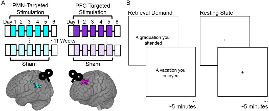
Experiment Design.
(A) Subjects received five consecutive daily sessions of high-frequency (20 Hz) repetitive TMS delivered to a subject-specific parietal cortex location of the PMN selected based on high resting-state fMRI connectivity with the hippocampus (PMN-targeted Stimulation). Subjects received real stimulation and sham stimulation during different weeks, in counterbalanced order with an average 11.5 week washout period between these conditions. Before and ~ 24 hr after stimulation, subjects completed fMRI and memory assessments (white boxes). The same procedures were performed for a distinct control group of subjects, but with stimulation delivered to subject-specific locations of out-of-network prefrontal cortex (PFC-Targeted Stimulation). Circles indicate stimulation locations for each participant. (B) fMRI connectivity was measured during the resting state and during an autobiographical memory retrieval task, for which subjects were shown prompts describing common life events and asked to vividly recall personal events matching the prompts.
Figure 2 with 1 supplement
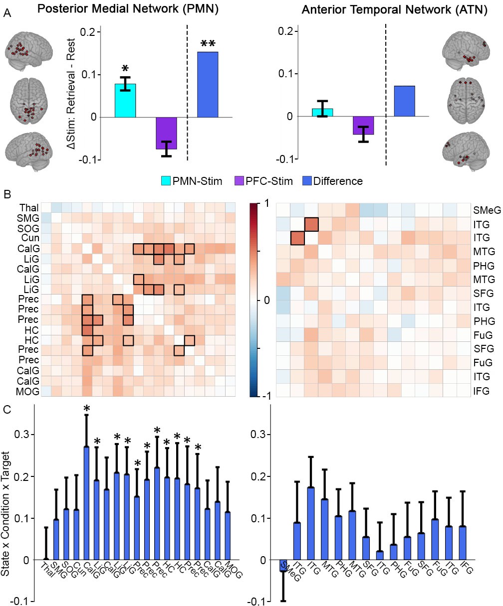
Greater memory task-dependent connectivity increases due to PMN-targeted versus PFC-targeted control stimulation for the PMN.
(A) Stimulation effect on mean connectivity (all regions to all other regions) during memory retrieval relative to rest for the PMN (left) and the ATN (right). Error bars indicate subject-level standard error of the mean. *=p < 0.05, **=p < 0.01 for the interaction between stimulation condition and state in each group, or the three-way interaction between condition, state, and stimulation location. (B) The same effect of stimulation on connectivity between each pair of regions in the PMN (left) or ATN (right). Coloration represents the beta-weight of the three-way interaction effect, with significant cells shown in bold (FDR corrected p<0.05). (C) The same effect of stimulation on connectivity for each region in the PMN to all other PMN regions (left) and each region in the ATN to all other ATN regions (right). Error bars indicate standard error of the mean of the three-way interaction. *=FDR corrected p<0.05. Regions are shown in Figure 2—figure supplement 1 and region abbreviations are expanded in Supplementary file 2.
-
Figure 2—source data 1
PMN/ATN network-based analysis.
Subject: 1–16 = PMN targeted group, 17–32 = PFC targeted group. State: Autobiographical memory task (Retrieval) or resting-state (Rest). Condition: Full stimulation (Stim) or sham stimulation (Sham). Target: PMN-targeted (Par) or PFC-targeted (Ant). Order: Full stimulation first (1) or sham stimulation first (2). Connectivity: Average activity correlation between network regions for that scan. tSNR: Average whole-brain temporal signal to noise ratio for that scan. Network: Posterior-medial (PM) or anterior-temporal (AT).
- https://doi.org/10.7554/eLife.49458.005
Figure 2—figure supplement 1

Network regions of interest.
Posterior medial network (PMN) and anterior temporal network (ATN) regions of interest (Ranganath and Ritchey, 2012): PMN (red), ATN (green).
Figure 3 with 3 supplements
Greater memory task-dependent connectivity increases due to PMN-targeted versus prefrontal-control stimulation.
(A) Regions showing a significant interaction between condition, task, and group, with red coloration indicating stimulation increased connectivity more during retrieval than during rest in the PMN-targeted group relative to the prefrontal-control group. All regions showed greater memory task-related connectivity change following PMN-targeted stimulation. Comprehensive view shown in Figure Supplement. (B) Mean stimulation effect on connectivity during memory retrieval relative to rest for all supra-threshold regions. Error bars are provided for illustrative purposes and indicate subject-level standard error of the mean for supra-threshold regions. Points indicate the mean effect for each supra-threshold region. Note: Statistical values are not indicated, as this would be redundant with the statistical definition of these supra-threshold regions.
-
Figure 3—source data 1
Whole-brain analysis, full model.
Subject: 1–16 = PMN targeted group, 17–32 = PFC targeted group. State: Autobiographical memory task (Retrieval) or resting-state (Rest). Condition: Full stimulation (Stim) or sham stimulation (Sham). Target: PMN-targeted (Parietal) or PFC-targeted (Prefrontal). Cluster: Significant cluster identified via whole-brain analysis. Memory: Percent of memory trials with correct context recollection. Connectivity: Average global correlation within the cluster for that scan. tSNR: Average whole-brain temporal signal to noise ratio for that scan.
- https://doi.org/10.7554/eLife.49458.010
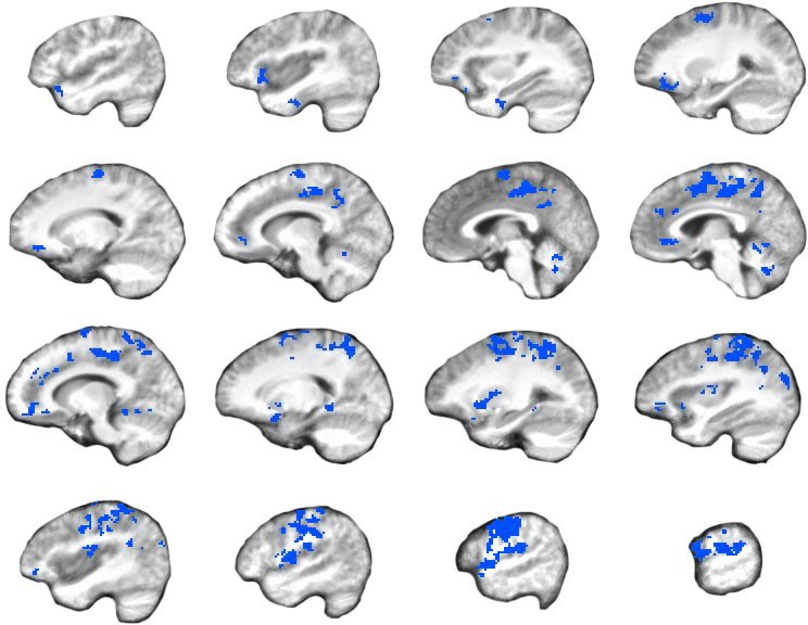
Figure 3—figure supplement 1
Selective effects of stimulation on memory-specific connectivity.
All regions showing a significant three-way interaction between condition, demand, and group. Red coloration indicates supra-threshold voxels with positive differences (retrieval stimulation effect > rest stimulation effect) and blue coloration indicates negative differences (rest stimulation effect > retrieval stimulation effect).
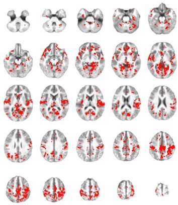
Figure 3—figure supplement 2
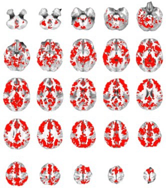
Increased fMRI connectivity during memory retrieval.
Regions showing a significant main effect of demand independent from stimulation condition or stimulation group. All showed greater connectivity during the retrieval demand relative to rest. Left = Left. Red coloration indicates supra-threshold voxels with positive differences (retrieval > rest).
Figure 3—figure supplement 3
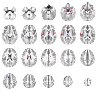
Common effects of stimulation across sites.
Regions showing a significant main effect of stimulation condition, regardless of demand or stimulation site. Left = Left. Red coloration indicates supra-threshold voxels with positive differences (stimulation >sham) and blue coloration indicates negative differences (sham > stimulation).
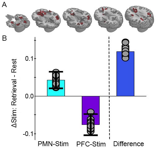
Figure 4 with 2 supplements
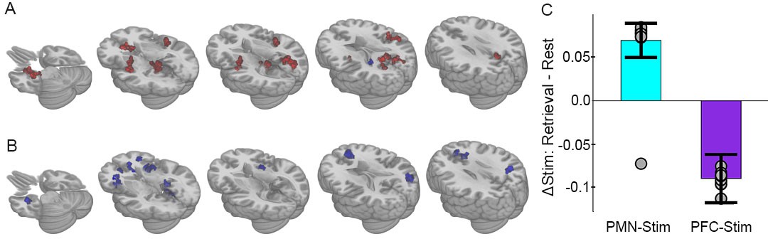
Selective effects of PMN-targeted stimulation on memory-related connectivity.
(A) Regions showing significant interaction between stimulation condition and cognitive task following PMN-targeted stimulation, with red coloration indicating stimulation increased connectivity more during retrieval than during rest and blue coloration indicating the opposite effect. Comprehensive view shown in Figure Supplements. (B) Regions showing significant interaction between stimulation condition and cognitive task following PFC-targeted control stimulation. (C) Mean stimulation effect on connectivity during memory retrieval relative to rest for all supra-threshold regions. Error bars are provided for illustrative purposes and indicate subject-level standard error of the mean for all supra-threshold regions. Points indicate the mean effect for each supra-threshold region. Note: Statistical values are not indicated, as this would be redundant with the statistical definition of these supra-threshold regions.
-
Figure 4—source data 1
Whole-brain analysis, PMN-targeted group.
Subject: 1–16 = PMN targeted group, 17–32 = PFC targeted group. State: Autobiographical memory task (Retrieval) or resting-state (Rest). Condition: Full stimulation (Stim) or sham stimulation (Sham). Cluster: Significant cluster identified via whole-brain analysis. Memory: Percent of memory trials with correct context recollection. Connectivity: Average global correlation within the cluster for that scan. tSNR: Average whole-brain temporal signal to noise ratio for that scan.
- https://doi.org/10.7554/eLife.49458.014
-
Figure 4—source data 2
Whole-brain analysis, PFC-targeted group.
Subject: 1-16=PMN-targeted group, 17-32=PFC-targeted group. State: Autobiographical memory task (Retrieval) or resting-state (Rest). Condition: Full stimulation (Stim) or sham stimulation (Sham). Cluster: Significant cluster identified via whole-brain analysis. Memory: Percent of memory trials with correct context recollection. Connectivity: Average global correlation within the cluster for that scan. tSNR: Average whole-brain temporal signal to noise ratio for that scan.
- https://doi.org/10.7554/eLife.49458.015
Figure 4—figure supplement 1

Selective effects of stimulation on memory-specific connectivity.
All regions showing a significant interaction between condition and demand following PMN-targeted stimulation. Red coloration indicates supra-threshold voxels with positive differences (retrieval stimulation effect > rest stimulation effect) and blue coloration indicates negative differences (rest stimulation effect > retrieval stimulation effect).
Figure 4—figure supplement 2
Selective effects of stimulation on memory-specific connectivity.
All regions showing a significant interaction between condition and demand following PFC-targeted stimulation. Red coloration indicates supra-threshold voxels with positive differences (retrieval stimulation effect > rest stimulation effect) and blue coloration indicates negative differences (rest stimulation effect > retrieval stimulation effect).
Figure 5
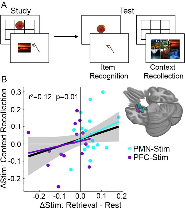
Increased memory-dependent connectivity predicts episodic memory improvement.
(A) Episodic memory task design. Participants studied trial-unique objects paired with either scene or location contexts. After a delay, we assessed object recognition memory and contextual recollection memory. (B) Connectivity values were pooled across the MTL regions identified in each group showing an interaction between stimulation condition and cognitive task. Scatterplot shows the relationship between each subject’s change in retrieval task connectivity relative to rest and their change in context recollection performance following stimulation. Greater specificity of connectivity change to the memory task in the left MTL was associated with improvement in context recollection in each group independently (cyan and purple regression lines) as well as collectively across all 32 subjects (black regression line).
Additional files
-
Supplementary file 1
Effect tables for the linear mixed models used to identify the demand-specific effects for PMN-targeted versus PFC-targeted control stimulation (Figure 1).
- https://doi.org/10.7554/eLife.49458.017
-
Supplementary file 2
Expanded names for PMN and ATN region abbreviations used in Figure 2.
Region labels from Eickhoff-Zilles macro labels from N27 in MNI space. Note that ‘calcarine gyrus’ refers to the area surrounding the calcarine sulcus, including the precuneus and lingual gyrus.
- https://doi.org/10.7554/eLife.49458.018
-
Supplementary file 3
18 supra-threshold regions identified via significant interaction between stimulation condition and demand (bolded), followed by their drivers—regions showing significant interaction between stimulation condition and demand in their seed-based connectivity to one of the 18 supra-threshold regions.
Region labels from Eickhoff-Zilles macro labels from N27 in MNI space. Note that ‘calcarine gyrus’ refers to the area surrounding the calcarine sulcus, including the precuneus and lingual gyrus.
- https://doi.org/10.7554/eLife.49458.019
-
Supplementary file 4
15 supra-threshold regions identified via significant interaction between stimulation condition and demand (bolded), followed by their drivers—regions showing significant interaction between stimulation condition and demand in their seed-based connectivity to one of the 15 supra-threshold regions.
Region labels from Eickhoff-Zilles macro labels from N27 in MNI space. Note that ‘calcarine gyrus’ refers to the area surrounding the calcarine sulcus, including the precuneus and lingual gyrus.
- https://doi.org/10.7554/eLife.49458.020
-
Transparent reporting form
- https://doi.org/10.7554/eLife.49458.021
Download links
A two-part list of links to download the article, or parts of the article, in various formats.
Downloads (link to download the article as PDF)
Open citations (links to open the citations from this article in various online reference manager services)
Cite this article (links to download the citations from this article in formats compatible with various reference manager tools)
Stimulating the hippocampal posterior-medial network enhances task-dependent connectivity and memory
eLife 8:e49458.
https://doi.org/10.7554/eLife.49458


